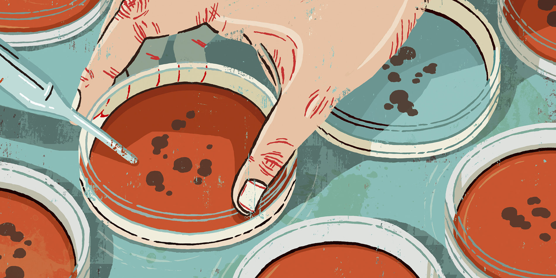
The fight against bacterial infections enters the 21st century
Millions of people die from bacterial infections every year, but the process of identifying bacteria is based on a century-old lab test.
Speeding up that process would save countless lives and is the goal of an interdisciplinary team of Stanford researchers.
The current “gold-standard” for identifying bacteria involves culturing samples – a process that is naturally slow and can span hours to days depending on the type of bacteria, says lead postdoctoral researcher and the project research co-director, Amr Saleh in the Department of Materials Science and Engineering at Stanford. Once that test is completed, it generally takes another 12–24 hours for doctors to determine which antibiotic is appropriate for treatment.
Because this process is so time-consuming, doctors tend to prescribe an antibiotic based on empiric guidelines that may or may not actually be suitable for a particular patient; the U.S. Centers for Disease Control and Prevention estimates that up to half of patients are unnecessarily treated with antibiotics or are treated with the wrong antibiotic type or dose. Incorrect use of antibiotics has led to the development of antibiotic-resistant bacteria, a significant global health threat. “Our interdisciplinary team of scientists, engineers, clinicians and doctors aims to expedite this process, rapidly and sensitively detecting bacteria in samples like blood and sputum,” says Jennifer Dionne, an associate professor of materials science and engineering and, by courtesy, of radiology.
The idea is to take the sample and then print it out into a two-dimensional array, where each pixel corresponds to a single cellular droplet. When the bacteria are illuminated with a particular color of light, their constituent molecules vibrate, changing the color of the scattered light. That color, or spectral shift, can be measured and used to uniquely identify the bacteria.
Using these techniques, the researchers were able to identify 31 species and strains of bacteria, representing 95% of all patient samples at Stanford Hospital, and the vast majority of all bacterial infections seen worldwide. Machine learning algorithms developed by the team facilitated rapid, accurate diagnosis, while low-cost hardware is enabling portable, point-of-care diagnostics.
More work needs to be done, but the results so far are promising: At this point, the average accuracy of the team’s approach in identifying the correct treatment of a bacterial infection is above 99%.
In addition to Dionne and Saleh, investigators on the team include Niaz Banaei, Stefano Ermon, Pierre Khuri-Yakub, Mark Holodniy, Manu Prakash, Sam Gambhir and Stefanie Jeffrey. Their work is funded by the Stanford Catalyst for Collaborative Solutions, an initiative launched in 2016 to inspire campus-wide collaborations to tackle some of the world’s most urgent challenges.
Transcript
Mark Horowitz: The last talk of the session will be on actually, one of the first projects that the Catalyst Program focused on. It was really a different way of looking at, and trying to analyze bacterial infections. So, it’s about a microbial cultural shift, and it’s gonna be presented by Jen Dionne and Amr Saleh, and they are gonna talk about this program that they’ve developed for basically being able to analyze infections.
Jen Dionne: Cool, well thank you so much for the introduction. Like Mark mentioned, my name is Jen Dionne, I’m a faculty member in material science and engineering. And I’m honored to present our work, which is in collaboration with a number of faculty spanning the schools of engineering, humanities and sciences, the school of medicine, and the school of business. And like Mark mentioned, we’re hoping to create a new diagnostic tool that can allow us to rapidly detect bacterial infections, as well as their antibiotic susceptibility.
I wanted to start off with a fun fact. There are close to 80 trillion cells in our bodies, and for every human cell that we have, there’s at least one bacterial cell. This microbiome is crucial for regulating metabolism, helping to aid in digestion, and to fight off infection.
But if an external bacterial cell comes along, it’s not always welcomed by our bodies with open arms. And if you look to the global statistics, you can see that bacterial infections have a profound impact on human health.
If we focus on just bacterial infections of the bloodstream, those bacterial bloodstream infections can cause more deaths than breast cancer, colon cancer, prostate cancer, and HIV combined in the United States. I think that’s a pretty staggering statistic. Also, if we look to developing countries, roughly about 30% of all deaths are due to bacterial infection, with a child dying every 20 seconds due to a bacterial related illness.
Why are these statistics so dire? It turns out that the gold standard for identifying bacterial infections is based on a century old culturing technique.
We take a sample, in our case, we’re focusing on bacterial bloodstream infections, we put it in a blood culture vial, and then you essentially just wait for the bacteria to grow and multiply until they become detectable by virtue of a pH change in the solution. And that can take anywhere from about 12 hours up to 40 days, depending on the type of bacterial infection that a person might have.
Then, once you’re done identifying bacteria in the sample, you have to determine which antibiotics it’s going to respond to. And that screening for antibiotic susceptibility is usually often done by taking the bacterial cultures, putting them on an agar plate with various concentrations of antibiotics, and various types, and just seeing where do the bacteria die. So, like I said, this test typically takes on the order of about two days. Meanwhile, your a patient in the hospital, and the doctor does not want you suffering, so is prescribing you either broad spectrum antibiotics, or kind of antibiotics based on empiric guidelines. And according to the CDC, roughly about half of all patients are either unnecessarily treated with antibiotics, or given the wrong type, or the wrong dose. And this is why antimicrobial resistance is becoming such a huge problem, and one that we’re certainly going to have to face within the coming decades.
Our approach is to expedite this process, potentially to the point where it’s even at the point of care, so that way we can rapidly, and sensitively detect bacteria in complex samples like blood, and mucus, and urine, and potentially even food and water.
And the idea is to take the sample, and then print it out into a two dimensional array, where each pixel corresponds to a single cellular droplet. And then, we essentially take a picture of this two dimensional array, where the spectrum, or the color of each pixel corresponds to the particular type of cell that’s in that pixel, as well as its species strain and antibiotic susceptibility. The nice thing is, it requires no sample specific preparation, or cell tagging, and it can also be strongly enhanced, so we can visualize the color with a number of tricks that our colleagues are developing.
For those of you who are familiar with bacteria, you probably know that they can be engineered to express different colors. There are even art competitions in the bay area, to make agar art with these engineered bacteria.
But for the most part, across the dozens or so of bacteria that you would run into, they’re colorless. So, how is it that based on this scattering spectrum, we can actually identify their color without having to do the color specific engineering?
Well, the idea is to use a technique called Raman scattering, and I’ll talk you through how it works. So here’s an example cell, just E.coli, and if we zoom in, it has a cell membrane that’s comprised of many different proteins. When we illuminate this with light, let’s say we’re illuminating it with green light, all of those proteins and molecules in that cell membrane are going to be vibrating. And those vibrations either add or subtract energy, to that incident green light. Then, we wind up getting say, red photons out, or blue photons out, and that’s something that we read out in what’s called a Raman scattering spectrum. Here we’re looking at the Raman shift, which is basically a measure of the energy that’s been added to or subtracted from the incident color of light that we put in. So, this Raman scattering provides a fingerprint of the bacteria, because each bacterial species and strain has a unique molecular composition that we can use for identification.
What we’ve done is look at single cells Raman spectra from 31 strains and 22 species, representing 95% of all patient samples at Stanford Hospital, and the vast majority of all bacterial infections seen worldwide. So, you can see, looking at these Raman spectra, that you can tell differences between different types of bacterial spectra, including staph, and pneumonia, and streptococcus.
But when you do a principal component analysis, and try to dive a little bit deeper into how you can separate out the different species and strains, you can see that on the left hand side of this plot, bacteria and blood, very unique, but all of the bacterial species and strains tend to cluster together. So, some are easily distinguishable, others are much more challenging to distinguish.
And this is where our group really relied on the expertise in the Bay Area, and in our computer science colleagues to develop a machine learning network that can help to classify all of the various Raman spectra, by using a convolutional neural network. So, this is the same sort of machine learning algorithm that you would use in image recognition, except now that we’re using it for spectral recognition.
What this algorithm does, is it looks at all of the spectra, and then based on the training set, it can take a new spectra and identify what species and strain it is, and it outputs a predictive score. So, given a new spectra, how likely is it to be one particular species or strain? So, here’s a subset of a so-called confusion matrix, where we have the true species on the column, and the predicted species across all of the various rows. And you can see for staph aureus and staph epidermis, we have fairly high classification accuracies.
If we average the classification accuracies across all species and strains, it’s about 82%, which is actually pretty good, given that we had some strains that were just isomorphs, or genetically engineered versions just to test how accurate our algorithm could be.
And what matters to a doctor is a first choice antibiotic treatment. So, if we look at what the antibiotic treatment groups would be, you can see that our average treatment accuracy is 97%, so this is better than many clinical asses that would be used. And then here, since I mentioned some of those genetically modified pairs that kind of brought down our accuracy, you can see those are more in the 80th percentile, but it’s still quite promising that we can just insert a resistance gene into a bacteria and also detect differences. So, this is very exciting to look at say, how bacteria might be evolving to be resistant to bacterium.
The final thing I’ll mention before handing off the clicker to my colleague, Amr, is that we’ve done preliminary clinical trials, where we looked at clinical samples. And in the first clinical trial corresponding to the 30 patients you see on the left, we did pretty good with the treatment groups, although we did have some misfires. But then, when we redid the experiment by using this clinical data set as training, we could improve the accuracy of our machine learning algorithm.
This is quite exciting, and you can see that we get above 99% accuracy when we start looking across all these patients. And importantly, we only need 10 bacterial cells to get accuracy exceeding 99% treatment accuracy.
Amr Saleh: So, now that we have seen that we can identify individual bacteria based on their signature or their optical scattering, the question is, how can we transfer this 3D, if we have a 3D volume of cells, how can we use this property to develop a diagnostic tool?
In a 3D volume like a blood sample, you have many, many different types of cells with different shapes and colors. If you try to identify them as they are in a vial, it’s impossible to detect their colors. But if you were to imagine that they can create a 2D paint out of this 3D volume, you’ll be able to identify the different colors of each of these cells. And this is exactly what we do, except that we don’t really do hand painting, but we do printing.
This is a schematic of how our tool works. We have our sample that we wanna analyze, mix it with some type of nanoparticles that make the optical signatures more vivid and easier to detect. And then, we use this printer to basically digitize this 3D volume into discrete pixels. Each of these pixels have an individual cell in a dispersion of nanoparticles. Then, we take images, take just simple pictures of these printouts, and use the colors from each of these individual pixels to measure the spectra, and generate the final report of if there is a pathogen in the sample, what type of pathogen, and what treatment should be used for this patient.
Let’s take a closer look at the printer. This is a real video for the printing process, where we have a focused acoustic waves, basically at the interface between the liquid and the air, and you can see how we can generate very controlled microdroplets with a very specific volume, based on the frequency of the transducers.
This is very advantageous when it comes handling vial samples, because you are controlling the volume that you’re ejecting with just waves, acoustic waves. You don’t need a physical nozzle or anything, so there is no clogging, and you are not killing the cells as they are being ejected. At the same time, you can also have a very precise control on the contents of the cell, and you can process large volumes of samples in a very short time.
This was our first trial with this printer, to eject, or to print individual droplets of E.coli into an agar plate, and you can see here how these individual droplets grew into colonies on the agar plate. But even more recently now, we can actually make up, or print more sophisticated words like Stanford. And this is actually written with a blood sample, a whole blood sample mixed with plasmonic nanoparticles. If you were to zoom in on that, you will see how these letters are actually formed from individual blood cells, printed. So, when we say that we can write Stanford name with our blood, it’s actually literally, we mean that.
Now that we have the sample printed out, how can we scan it? How can we screen it for infections in there? We have been using our scientific grade tools, which are heavy, expensive, bulky, and they’re by no means, portable. But our goal is to make a point of care, low-cost device. So, we tackled this challenge in two ways.
First, we ask ourselves, these are the data that sophisticated, high end tools give in our lab, so do we need all the features? Do we need all the spectral details in here, so that we can make accurate identification?
Turns out, that we don’t really need this. Only certain bands, and certain colors, or certain wave lengths in this spectrum, matters for us. And if you only take these bands, or these barcodes, we find that the classification accuracy can be actually 95%, just by using these selected features. And we are still optimizing that to get this to even higher than the 95%. The other strategy, the complimentary strategy now, is that knowing that we don’t need all the spectral resolution, or all the details in the spectra, we can design our own hardware that is optimized for this specific application.
This is what we did — we’re actually using commercial CMOS camera that is made for machine vision applications, with a transmission grading, to build our home built spectrometer. And this is the physical implementation of that, and if you can see in this picture, this is a handheld. This is a compact, light device that you can hold in your hand, and it cost us less than $500, and this is the first spectra that we measured with this new device. It was a really exciting moment to see this really nice spectra, with just very cheap spectrometer that we built.
Where we are right now, we are working on integrating these different components together in one standalone platform, where we can have a blood sample, and give us at the end, a report in less than an hour. And we aim to test this integrated platform with the Stanford Hospital and the VA Hospital, using real patient samples, and hopefully run some clinical trials in the field, where the resources are very strict.
To conclude this talk, we have shown you today that we can identify individual bacteria solely from their optical signatures, and we can digitize a 3D volume of a sophisticated cellular sample using a bio-printer to make it easy to use a low-cost spectrometer, or a camera to identify the bacteria. And finally, I would like to thank all our amazing team members who made this work possible, and of course the Catalyst funds, who actually brought us together and made all this possible.
And thank you all for coming in, thank you.


